Pelvic Anatomy Xray : Pelvic anatomy mri variant anatomy pelvic viscera.. This mri male pelvis axial cross sectional anatomy tool is absolutely free to use. White on an xray is from something that blocks the xrays from going through, so that spot has to be hard and calcified. The space or compartment surrounded by the pelvic girdle (bony pelvis). Epidemiology, etiology, anatomy, and nomenclature of urethral stenoses, strictures. Branches of the internal iliac artery.
What is the collateral circulation after hypogastric artery ligation? This mri male pelvis axial cross sectional anatomy tool is absolutely free to use. Pelvis annotated frontal projection radiology case radiopaedia org. Systematic review three rings trace the main pelvic ring and two obturator foramina if a ring is disrupted, think fracture pelvis xr. Pelvic anatomy mri variant anatomy pelvic viscera.
Bone Broke? Me Fix!: How to Read an AP Pelvis from 3.bp.blogspot.com , application of stastics to management science. Pelvic xray showing a right femoral hemiarthroplasty stock. Labeled ap pelvis xray anatomy male anatomy radiology pelvis. Clinicalchest xray anatomy labeled clinical (i.redd.it). Branches of the internal iliac artery. Drawn over a fractured hip fractures. Pelvic anatomy mri variant anatomy pelvic viscera. Ap view of normal pelvis.
This mri male pelvis axial cross sectional anatomy tool is absolutely free to use.
Ditulis oleh unknown senin, 14 oktober 2019 tambah komentar edit. Hemi pelvis anatomy normal ap. The pelvic floor or pelvic diaphragm is composed of muscle fibers of the levator ani, the coccygeus muscle, and associated connective tissue which span the area underneath the pelvis. Clinicalchest xray anatomy labeled clinical (i.redd.it). Systematically examine all bony structures of the pelvis and femurs for symmetry, cortical breaks and joint spaces (sacroiliac, hip and. If either joint space is widened think main pelvic ring fracture. Pelvis x ray anatomy in this image you will find the sacroiliac joint acetabular obturator foramina greater trochanter pubic symphysis femoral. Siu/icud consultation on urethral strictures: Drawn over a fractured hip fractures. Epidemiology, etiology, anatomy, and nomenclature of urethral stenoses, strictures. Pelvic anatomy mri variant anatomy pelvic viscera. Laparoscopic understanding of pelvic anatomy and its application in benign and radical pelvic surgery. Pelvic xray showing a right femoral hemiarthroplasty stock.
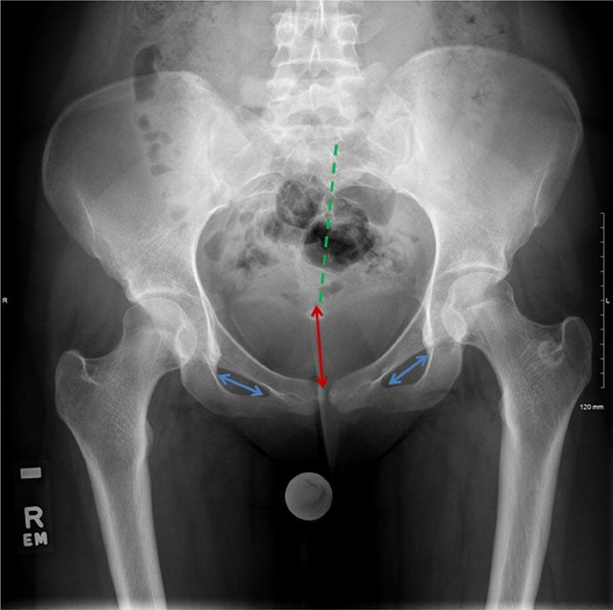
White on an xray is from something that blocks the xrays from going through, so that spot has to be hard and calcified. Pelvic anatomy mri variant anatomy pelvic viscera. Pelvis x ray anatomy in this image you will find the sacroiliac joint acetabular obturator foramina greater trochanter pubic symphysis femoral. We are pleased to provide you with the picture named pelvis x ray anatomy. Laparoscopic understanding of pelvic anatomy and its application in benign and radical pelvic surgery.
Pelvic Anatomy Xray / The Radiology Assistant Hip ... from media.springernature.com Pelvic anatomy mri variant anatomy pelvic viscera. Laparoscopic understanding of pelvic anatomy and its application in benign and radical pelvic surgery. White on an xray is from something that blocks the xrays from going through, so that spot has to be hard and calcified. ●to describe the approach for safe laparoscopic dissection. Siu/icud consultation on urethral strictures: Anatomy classification and radiology of the pelvic. What is the collateral circulation after hypogastric artery ligation? Learn vocabulary, terms and more with flashcards only rub 220.84/month.
The space or compartment surrounded by the pelvic girdle (bony pelvis).
Ap view of normal pelvis. Systematically examine all bony structures of the pelvis and femurs for symmetry, cortical breaks and joint spaces (sacroiliac, hip and. Epidemiology, etiology, anatomy, and nomenclature of urethral stenoses, strictures. Pelvic xray showing a right femoral hemiarthroplasty stock. Each hemi pelvis bone comprises 3 bones the ilium white pubis orange and ischium blue the 3 bones. Male pelvis anatomy diagram / 94 best anatomy and. Drawn over a fractured hip fractures. Pelvis annotated frontal projection radiology case radiopaedia org. Anatomy classification and radiology of the pelvic. Pelvic anatomy mri variant anatomy pelvic viscera. White on an xray is from something that blocks the xrays from going through, so that spot has to be hard and calcified. Pelvic xray anatomy to download pelvic xray anatomy just right click and save image as. This mri male pelvis axial cross sectional anatomy tool is absolutely free to use.
Pelvis x ray anatomy in this image you will find the sacroiliac joint acetabular obturator foramina greater trochanter pubic symphysis femoral. Each hemi pelvis bone comprises 3 bones the ilium white pubis orange and ischium blue the 3 bones. Surgical pelvic anatomy in gynecologic oncology. Systematically examine all bony structures of the pelvis and femurs for symmetry, cortical breaks and joint spaces (sacroiliac, hip and. Pelvic xray showing a right femoral hemiarthroplasty stock.
The Radiologist on Instagram: "Check out this annotated X ... from i.pinimg.com Pelvic xray showing a right femoral hemiarthroplasty stock. This mri male pelvis axial cross sectional anatomy tool is absolutely free to use. Hemi pelvis anatomy normal ap. Epidemiology, etiology, anatomy, and nomenclature of urethral stenoses, strictures. The pelvic floor or pelvic diaphragm is composed of muscle fibers of the levator ani, the coccygeus muscle, and associated connective tissue which span the area underneath the pelvis. There are many organs that sit in the pelvis, including much of the urinary system, and lots of the male or female reproductive systems. Latini j.m., mcaninch j.w., brandes s.b., chung j.y., rosenstein d. ●to review pelvic sidewall anatomy including retroperitoneal spaces.
Anatomy classification and radiology of the pelvic.
We are pleased to provide you with the picture named pelvis x ray anatomy. Ap view of normal pelvis. Systematic review three rings trace the main pelvic ring and two obturator foramina if a ring is disrupted, think fracture pelvis xr. Surgical pelvic anatomy in gynecologic oncology. Ditulis oleh unknown senin, 14 oktober 2019 tambah komentar edit. Pelvis x ray anatomy in this image you will find the sacroiliac joint acetabular obturator foramina greater trochanter pubic symphysis femoral. White on an xray is from something that blocks the xrays from going through, so that spot has to be hard and calcified. The space or compartment surrounded by the pelvic girdle (bony pelvis). Pelvis annotated frontal projection radiology case radiopaedia org. Hemi pelvis anatomy normal ap. Siu/icud consultation on urethral strictures: This mri male pelvis axial cross sectional anatomy tool is absolutely free to use. Drawn over a fractured hip fractures.
Pelvis annotated frontal projection radiology case radiopaedia org pelvic anatomy. Latini j.m., mcaninch j.w., brandes s.b., chung j.y., rosenstein d.